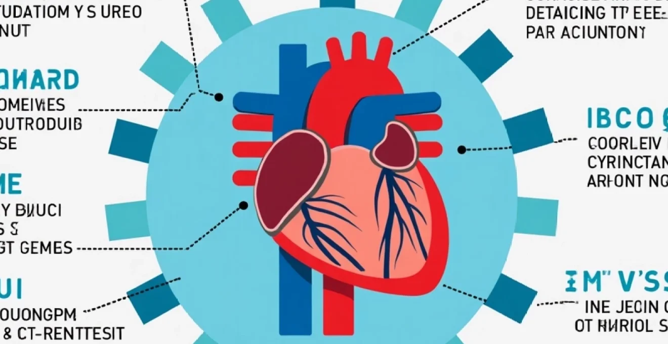
Heart disease remains a leading cause of mortality worldwide, but early detection can significantly improve outcomes. Understanding the available screening options is crucial for maintaining cardiovascular health. From basic risk assessments to advanced imaging techniques, a wide array of tests can help identify potential heart problems before they become life-threatening. This comprehensive guide explores the most effective screening methods, their applications, and when you should consider asking your healthcare provider about them.
Cardiovascular risk assessment: primary screening methods
The first step in heart disease screening often involves assessing your overall cardiovascular risk. Several well-established tools can help healthcare providers estimate your likelihood of developing heart disease in the coming years. These assessments consider various factors, including age, gender, blood pressure, cholesterol levels, and lifestyle habits.
Framingham risk score: calculating 10-year CVD risk
The Framingham Risk Score is one of the most widely used cardiovascular risk assessment tools. Developed from the long-running Framingham Heart Study, this algorithm estimates your 10-year risk of developing cardiovascular disease (CVD). The score takes into account factors such as age, gender, total cholesterol, HDL cholesterol, smoking status, and systolic blood pressure.
Healthcare providers use the Framingham Risk Score to categorise patients into low, intermediate, or high-risk groups. This classification helps determine the appropriate level of preventive care and whether additional testing is necessary. For example, individuals with a high Framingham score might be advised to undergo more intensive screening procedures.
Reynolds risk score: integrating C-Reactive protein levels
The Reynolds Risk Score is another valuable tool for assessing cardiovascular risk. What sets this score apart is its inclusion of high-sensitivity C-reactive protein (hs-CRP) levels, a marker of inflammation associated with increased cardiovascular risk. By incorporating hs-CRP, the Reynolds Risk Score may provide a more comprehensive assessment, especially for individuals without traditional risk factors.
This score is particularly useful for women and can help identify those who might benefit from preventive measures despite having normal cholesterol levels. The Reynolds Risk Score exemplifies how newer biomarkers can enhance our ability to predict and prevent heart disease.
QRISK3 algorithm: UK-Specific heart disease prediction
For those in the United Kingdom, the QRISK3 algorithm offers a tailored approach to cardiovascular risk assessment. This tool was developed using data from UK primary care patients and takes into account additional factors such as ethnicity, body mass index, and family history of heart disease. The QRISK3 provides a more accurate risk prediction for the UK population compared to other international models.
Healthcare providers in the UK often use QRISK3 to guide decisions about preventive treatments, such as statins. The algorithm’s ability to account for regional and ethnic variations makes it an essential tool in personalised cardiovascular care.
Coronary artery calcium (CAC) scoring: quantifying arterial calcification
Coronary Artery Calcium (CAC) scoring is a more advanced screening method that uses computed tomography (CT) to detect and quantify calcium deposits in the coronary arteries. These calcium deposits are indicators of atherosclerosis, the buildup of plaque that can lead to heart attacks and strokes.
CAC scoring is particularly valuable for individuals with intermediate risk based on traditional assessments. A high CAC score can reclassify a patient to a higher risk category, prompting more aggressive preventive measures. Conversely, a CAC score of zero in an intermediate-risk individual might suggest a lower actual risk, potentially influencing treatment decisions.
Non-invasive diagnostic tests for heart disease
Once initial risk assessments indicate a need for further evaluation, several non-invasive tests can provide detailed information about heart structure and function. These tests are crucial for diagnosing specific heart conditions and guiding treatment decisions.
Electrocardiogram (ECG): detecting electrical abnormalities
An electrocardiogram, or ECG, is a fundamental test in cardiac diagnostics. This simple, painless procedure records the heart’s electrical activity, providing valuable information about heart rate, rhythm, and conduction. ECGs can detect a wide range of heart abnormalities, from arrhythmias to signs of previous heart attacks.
Healthcare providers often use ECGs as a first-line diagnostic tool when patients present with symptoms such as chest pain, palpitations, or shortness of breath. Regular ECG screening can also be beneficial for individuals with known risk factors or a family history of heart disease.
Echocardiogram: assessing heart structure and function
An echocardiogram uses ultrasound waves to create detailed images of the heart’s structure and function. This test can reveal information about heart chamber size, wall thickness, valve function, and the heart’s pumping efficiency. Echocardiograms are invaluable for diagnosing conditions such as heart valve disorders, cardiomyopathies, and congenital heart defects.
There are several types of echocardiograms, including transthoracic (the most common), transesophageal, and stress echocardiograms. Each type has specific applications and can provide unique insights into cardiac health. For instance, stress echocardiograms can help identify coronary artery disease by showing how the heart responds to physical exertion.
Exercise stress test: evaluating cardiovascular performance
Exercise stress tests, also known as treadmill tests or stress ECGs, assess how the heart responds to physical exertion. During this test, patients exercise on a treadmill or stationary bicycle while their heart rate, blood pressure, and ECG are monitored. The test can reveal problems with blood flow to the heart that may not be apparent when the body is at rest.
Stress tests are particularly useful for diagnosing coronary artery disease and determining safe levels of physical activity for patients with known heart conditions. They can also help evaluate the effectiveness of treatments and guide rehabilitation programs for heart patients.
Cardiac CT angiography: visualising coronary artery blockages
Cardiac CT angiography is an advanced imaging technique that provides detailed pictures of the coronary arteries. This non-invasive test uses a special dye and CT scanning to create 3D images of the heart and blood vessels. It’s particularly effective at detecting narrowed or blocked coronary arteries, which can cause chest pain and heart attacks.
CT angiography is often recommended for patients with intermediate risk of coronary artery disease or those with inconclusive results from other tests. Its ability to visualise soft plaque in the arteries makes it a valuable tool for early detection of heart disease, even before symptoms appear.
Cardiac MRI: advanced imaging for structural heart defects
Cardiac Magnetic Resonance Imaging (MRI) provides highly detailed images of the heart’s structure and function without using radiation. This test is particularly useful for evaluating complex heart conditions, such as congenital heart defects, cardiomyopathies, and heart valve disorders. Cardiac MRI can also assess the extent of damage after a heart attack and help guide treatment decisions.
The exceptional image quality and ability to capture the heart in motion make cardiac MRI an invaluable tool in modern cardiology. It’s often used when other imaging methods are inconclusive or when a more comprehensive evaluation of heart function is needed.
Blood tests for cardiovascular health assessment
Blood tests play a crucial role in assessing cardiovascular health and identifying risk factors for heart disease. These tests can provide valuable information about cholesterol levels, inflammation markers, and specific proteins associated with heart damage.
Lipid panel: measuring cholesterol and triglyceride levels
A lipid panel is a group of tests that measure different types of lipids (fats) in your blood, including total cholesterol, low-density lipoprotein (LDL) cholesterol, high-density lipoprotein (HDL) cholesterol, and triglycerides. High levels of LDL cholesterol and triglycerides, along with low levels of HDL cholesterol, are associated with an increased risk of heart disease and stroke.
Regular lipid panel testing is recommended for adults, with the frequency depending on age, risk factors, and previous results. The results of a lipid panel can guide decisions about lifestyle changes and medications to reduce cardiovascular risk.
High-sensitivity C-Reactive protein (hs-CRP): inflammatory marker analysis
High-sensitivity C-reactive protein (hs-CRP) is a marker of inflammation in the body. Elevated levels of hs-CRP have been associated with an increased risk of heart disease, even in individuals with normal cholesterol levels. This test can provide additional information about cardiovascular risk, particularly when combined with traditional risk factors.
The hs-CRP test is often recommended for individuals at intermediate risk of heart disease based on other factors. Results can help healthcare providers make decisions about preventive treatments, such as statins, especially in cases where the need for treatment is not clear based on cholesterol levels alone.
Brain natriuretic peptide (BNP): detecting heart failure
Brain Natriuretic Peptide (BNP) is a hormone released by the heart in response to increased pressure or stretching of the heart muscle. Elevated levels of BNP in the blood can indicate heart failure or other conditions that put stress on the heart. The BNP test is particularly useful in diagnosing and monitoring heart failure.
Healthcare providers often use BNP levels to assess the severity of heart failure and monitor the effectiveness of treatment. This test can also help differentiate between heart failure and other conditions that cause similar symptoms, such as lung problems.
Troponin test: identifying myocardial infarction
Troponin is a protein released into the bloodstream when heart muscle is damaged. A troponin test is primarily used to diagnose heart attacks (myocardial infarction). Elevated troponin levels can indicate heart muscle damage, even when other signs of a heart attack are not present.
In emergency settings, troponin tests are crucial for quickly identifying patients experiencing a heart attack. However, troponin levels can also be elevated in other conditions that affect the heart, such as myocarditis or certain types of cardiomyopathy.
Genetic testing for hereditary heart conditions
Genetic testing has become an increasingly important tool in identifying individuals at risk for inherited heart conditions. These tests can reveal genetic mutations associated with various cardiac disorders, allowing for early intervention and family screening.
Familial hypercholesterolemia (FH) gene panel
Familial Hypercholesterolemia (FH) is an inherited condition characterised by very high levels of LDL cholesterol from birth. Genetic testing for FH involves analysing specific genes, such as LDLR, APOB, and PCSK9, which are known to be associated with the condition. Identifying FH through genetic testing is crucial because individuals with this condition have a significantly higher risk of early-onset heart disease.
Early diagnosis of FH through genetic testing allows for aggressive treatment to lower cholesterol levels and reduce the risk of heart attacks. It also enables cascade screening of family members who may be affected but undiagnosed.
Long QT syndrome (LQTS) genetic screening
Long QT Syndrome (LQTS) is a heart rhythm disorder that can cause fast, chaotic heartbeats, potentially leading to fainting spells or sudden cardiac arrest. Genetic testing for LQTS involves analysing several genes associated with the condition, including KCNQ1, KCNH2, and SCN5A.
Identifying LQTS through genetic testing is vital because the condition can be life-threatening if untreated. Early diagnosis allows for preventive measures, such as avoiding certain medications and lifestyle modifications. It also informs decisions about treatments like beta-blockers or implantable cardioverter-defibrillators (ICDs).
Hypertrophic cardiomyopathy (HCM) gene analysis
Hypertrophic Cardiomyopathy (HCM) is a condition where the heart muscle becomes abnormally thick, making it harder for the heart to pump blood. Genetic testing for HCM typically involves analysing genes such as MYH7, MYBPC3, and TNNT2, among others. HCM is a common cause of sudden cardiac death in young athletes, making early detection crucial.
Genetic testing for HCM can identify individuals at risk before they develop symptoms or structural changes in the heart. This allows for close monitoring, lifestyle adjustments, and in some cases, preventive treatments to reduce the risk of complications.
Age-specific cardiovascular screening recommendations
Cardiovascular screening needs change throughout life, with different tests and assessments becoming more relevant at various ages. Understanding age-specific recommendations can help ensure appropriate and timely screening for heart disease.
Young adults (20-39): establishing baseline cardiovascular health
For young adults, the focus is on establishing baseline measurements and identifying early risk factors. Key screenings for this age group include:
- Blood pressure checks at least every 2 years
- Lipid panel starting at age 20, then every 4-6 years if results are normal
- Body Mass Index (BMI) calculation at regular check-ups
- Assessment of smoking status and counselling on smoking cessation if needed
- Evaluation of physical activity levels and diet
Young adults with a family history of early heart disease or genetic conditions may require more frequent or additional screenings. It’s also important to discuss any symptoms or concerns with a healthcare provider, even at this young age.
Middle-aged adults (40-64): intensified risk factor assessment
As individuals enter middle age, the risk of heart disease increases, necessitating more comprehensive screening. Recommendations for this age group include:
- Annual blood pressure checks
- Lipid panel every 1-2 years, or more frequently if abnormal
- Diabetes screening, especially for those with risk factors
- Calculation of 10-year cardiovascular risk using tools like QRISK3 or Framingham Risk Score
- Consideration of advanced tests like CAC scoring for those at intermediate risk
Middle-aged adults should also discuss their individual risk factors with their healthcare provider to determine if additional tests, such as stress tests or echocardiograms, are warranted.
Older adults (65+): comprehensive cardiovascular evaluation
For older adults, cardiovascular screening becomes even more crucial, as the risk of heart disease significantly increases with age. Recommended screenings include:
- Blood pressure checks at every healthcare visit
- Annual lipid panel, unless otherwise indicated
- Regular assessment of cardiovascular symptoms
- Evaluation of cognitive function and its potential impact on cardiovascular health
- Consideration of functional capacity and frailty in treatment decisions
Older adults may also benefit from more frequent ECGs and may require adjustments to screening intervals based on overall health status and life expectancy.
Emerging technologies in heart disease screening
The field of cardiovascular screening is constantly evolving, with new technologies offering the potential for more accurate, efficient, and accessible heart disease detection. These emerging tools are shaping the future of cardiac diagnostics and preventive care.
Artificial intelligence in ECG interpretation: DeepHeart algorithm
Artificial Intelligence (AI) is revolutionising ECG interpretation, enhancing both speed and accuracy. The DeepHeart algorithm, for example, uses deep learning to analyse ECG data and detect subtle patterns that might be missed by human interpreters. This AI-powered approach can identify signs of conditions like atrial fibrillation, left ventricular hypertrophy, and even low ejection fraction from a standard ECG.
The integration of AI into ECG analysis has the potential to improve early detection of heart disease, especially in primary care settings where specialist interpretation might not be immediately available
. The potential for AI to enhance ECG interpretation could lead to more widespread and effective screening for heart disease, particularly in underserved areas or resource-limited settings.
Wearable devices: continuous heart rhythm monitoring
Wearable devices like smartwatches and fitness trackers are increasingly incorporating advanced heart monitoring features. These devices can provide continuous heart rate monitoring and even detect irregular heart rhythms, potentially identifying conditions like atrial fibrillation before they cause symptoms.
The Apple Watch, for example, includes an ECG app that can record an electrocardiogram and detect atrial fibrillation. Other devices offer features like blood oxygen monitoring and stress tracking, which can provide valuable data about overall cardiovascular health. While not a replacement for medical-grade devices, these consumer wearables can serve as an early warning system, prompting users to seek medical attention when abnormalities are detected.
Liquid biopsy: detecting circulating biomarkers for CVD
Liquid biopsy, a technique traditionally used in cancer diagnostics, is now being explored for cardiovascular disease screening. This approach involves analysing blood samples for circulating biomarkers, including cell-free DNA, microRNAs, and extracellular vesicles, which can indicate cardiovascular health status.
Research is ongoing to identify specific biomarkers that could reliably predict the onset or progression of heart disease. The potential advantages of liquid biopsy include its non-invasive nature and the ability to provide a comprehensive picture of cardiovascular health from a simple blood draw. As this technology develops, it could offer a powerful new tool for early detection and monitoring of heart disease.
3D printing in cardiovascular imaging: patient-specific heart models
3D printing technology is making significant strides in cardiovascular imaging and treatment planning. By creating detailed, patient-specific heart models based on CT or MRI scans, doctors can gain a better understanding of complex cardiac anatomy and plan interventions more effectively.
These 3D-printed models are particularly valuable for planning surgeries for congenital heart defects or complex valve repairs. They allow surgeons to visualise and practice procedures before entering the operating room, potentially improving outcomes and reducing surgical time. Additionally, these models can be used for patient education, helping individuals better understand their condition and proposed treatments.
As 3D printing technology becomes more refined and accessible, it has the potential to become a standard tool in cardiovascular care, enhancing both diagnosis and treatment planning for a wide range of heart conditions.
HTML