Discovering small white bumps on your lips can be both alarming and perplexing, particularly when they appear without warning or obvious cause. These minute lesions represent a diverse spectrum of dermatological conditions, ranging from benign sebaceous gland variations to more complex inflammatory processes requiring professional intervention. Understanding the underlying mechanisms behind these formations is crucial for appropriate management and peace of mind.
The delicate vermillion border of the lips provides a unique anatomical landscape where various skin conditions manifest differently than on other body regions. This specialised tissue, rich in blood vessels and nerve endings, creates an environment where normal physiological processes can sometimes produce visible abnormalities. The interplay between hormonal fluctuations, genetic predisposition, and environmental factors contributes to the development of these characteristic white lesions.
Modern dermatological research has revealed that what appears as simple “white bumps” actually encompasses several distinct pathological processes, each requiring specific diagnostic approaches and treatment protocols. The prevalence of these conditions varies significantly across different age groups and populations, with some manifestations being nearly universal whilst others remain relatively rare. Recognition of these patterns enables both healthcare professionals and individuals to make informed decisions about when intervention is necessary .
Fordyce spots: sebaceous gland hyperplasia on lip vermillion
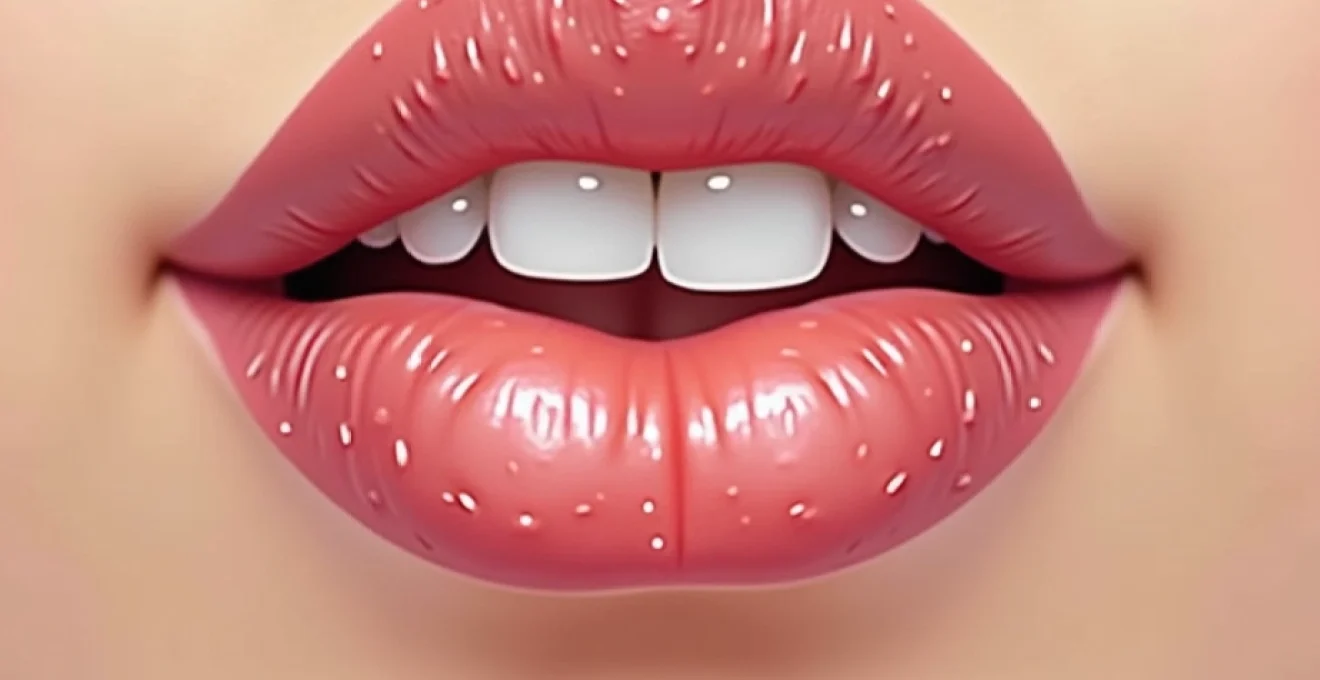
Fordyce spots represent one of the most commonly encountered forms of white lip bumps, affecting an estimated 70-80% of the adult population to varying degrees. These lesions manifest as clusters of small, pale yellow or white papules, typically measuring 1-3 millimetres in diameter, distributed along the vermillion border and extending onto the labial mucosa. The condition represents a hyperplastic response of ectopic sebaceous glands, which exist naturally within the oral cavity but become visually prominent under specific circumstances.
The pathophysiology underlying Fordyce spot development involves the enlargement and increased activity of sebaceous glands that are not associated with hair follicles. Unlike conventional sebaceous glands found elsewhere on the body, these ectopic structures lack the typical pilosebaceous unit architecture. This anatomical variation predisposes them to becoming more visible during periods of hormonal fluctuation , particularly during adolescence when androgenic stimulation peaks.
Ectopic sebaceous gland distribution patterns
The distribution of ectopic sebaceous glands throughout the oral cavity follows predictable anatomical patterns, with the highest concentration observed along the lateral aspects of the lips and the buccal mucosa. Research indicates that approximately 95% of individuals possess these glands, though their visibility varies considerably based on individual factors including skin thickness, pigmentation, and hormonal status. The glands demonstrate bilateral symmetry in most cases, creating the characteristic paired appearance often observed in clinical presentations.
Histopathological characteristics of fordyce granules
Microscopic examination of Fordyce spots reveals enlarged sebaceous lobules composed of mature sebocytes arranged around central ducts. The histological architecture closely resembles that of conventional sebaceous glands, with peripheral basal cells gradually differentiating into lipid-laden sebocytes towards the centre. The absence of inflammatory infiltrate distinguishes these lesions from pathological conditions requiring therapeutic intervention . Immunohistochemical staining demonstrates positive reactions for sebaceous differentiation markers, confirming their benign nature.
Differential diagnosis from molluscum contagiosum
Clinical differentiation between Fordyce spots and molluscum contagiosum lesions requires careful attention to morphological details and distribution patterns. Molluscum contagiosum typically presents as umbilicated papules with a characteristic central depression, whilst Fordyce spots maintain a smooth, dome-shaped surface without central umbilication. Additionally, molluscum lesions tend to occur as isolated papules or small clusters, whereas Fordyce spots characteristically appear in linear arrangements along the lip border.
Hormonal influences on sebaceous gland proliferation
Androgenic hormones play a pivotal role in sebaceous gland development and activity throughout life, with testosterone and its metabolite dihydrotestosterone serving as primary stimulatory factors. During puberty, increased androgen production leads to sebaceous gland enlargement and enhanced sebum production, often coinciding with the initial appearance or increased visibility of Fordyce spots. This hormonal influence explains why these lesions become more prominent during adolescence and may fluctuate with hormonal cycles throughout adulthood .
The relationship between hormonal status and Fordyce spot prominence underscores the importance of considering endocrine factors when evaluating lip lesions in adolescent and adult populations.
Milia formation and keratin retention cysts
Milia represent another prevalent cause of white lip bumps, characterised by small, firm, white or yellow papules resulting from keratin entrapment within miniature epidermal cysts. These lesions typically measure 1-2 millimetres in diameter and demonstrate a characteristic pearly appearance under magnification. Unlike Fordyce spots, milia exhibit a harder consistency upon palpation and may persist for extended periods without spontaneous resolution.
The pathogenesis of milia involves the abnormal retention of keratinocytes and cornified material within dilated pilosebaceous structures or eccrine ducts. This process creates microscopic cysts lined by stratified squamous epithelium, filled with laminated keratin debris. The condition can occur spontaneously or develop secondary to various predisposing factors, including trauma, inflammatory processes, or the use of occlusive topical agents.
Primary milia development mechanisms
Primary milia arise spontaneously without identifiable precipitating factors, representing a developmental variation in epithelial differentiation and desquamation processes. These lesions typically appear during infancy or childhood, affecting approximately 40-50% of newborns before resolving spontaneously within several weeks. In adults, primary milia may develop de novo, particularly in individuals with genetic predispositions affecting epithelial cell turnover rates.
Secondary milia following lip trauma
Secondary milia formation occurs as a consequence of various traumatic insults to the lip region, including thermal burns, chemical injuries, surgical procedures, or chronic irritation from dental appliances. The healing process following such trauma can disrupt normal epithelial architecture, creating conditions favourable for keratin entrapment. Recognition of this relationship is crucial for patients with a history of lip trauma who subsequently develop persistent white bumps .
Epidermal inclusion cyst pathogenesis
The development of epidermal inclusion cysts shares common pathophysiological mechanisms with milia formation, though these lesions typically achieve larger dimensions and demonstrate deeper dermal involvement. The process begins with the implantation or invagination of surface epithelium into deeper tissue layers, where continued keratinisation creates expanding cystic structures. These cysts maintain connection to surface epithelium through narrow channels, distinguishing them from true dermal cysts.
Viral aetiology: human papillomavirus and herpes simplex
Viral infections represent a significant category of lip lesions that may initially present as white or flesh-coloured bumps before developing their characteristic features. The oral cavity’s exposure to environmental pathogens, combined with the rich vascular supply and mucosal surfaces of the lips, creates an environment conducive to viral replication and transmission. Understanding the viral causes of lip bumps is essential for appropriate diagnosis, treatment, and prevention of transmission to others.
The most clinically relevant viral pathogens affecting the lip region include human papillomavirus (HPV), herpes simplex virus (HSV), molluscum contagiosum virus, and occasionally Epstein-Barr virus. Each of these pathogens demonstrates distinct tropism for specific cell types and anatomical locations, resulting in characteristic clinical presentations that aid in differential diagnosis. Early recognition of viral aetiology enables prompt initiation of appropriate antiviral therapy and implementation of infection control measures .
HPV-6 and HPV-11 associated oral papillomas
Human papillomavirus types 6 and 11 are the predominant causative agents of benign oral papillomas affecting the lip region. These low-risk HPV types demonstrate particular affinity for mucosal surfaces, where they induce benign epithelial proliferation resulting in characteristic papillomatous growths. Initial lesions may appear as small white or pink bumps before developing the classic “cauliflower-like” surface texture associated with mature papillomas.
The viral lifecycle involves infection of basal epithelial cells, where HPV DNA integrates into the host cell genome and directs the production of viral proteins that stimulate cell proliferation. This process typically requires several months to produce clinically visible lesions , explaining the often delayed appearance of papillomas following initial exposure. Treatment approaches focus on physical destruction of infected tissue combined with immune system enhancement to prevent recurrence.
HSV-1 vesicular eruptions and healing phases
Herpes simplex virus type 1 infections of the lip region follow a characteristic clinical course, beginning with prodromal symptoms including tingling, burning, or numbness, followed by the rapid development of clustered vesicles. During the initial phase, these vesicles may appear as small white or clear bumps filled with viral-laden fluid. The progression from vesicles to pustules, then to crusted lesions, typically occurs over 7-10 days in immunocompetent individuals.
The viral replication cycle involves initial infection of epithelial cells, followed by retrograde axonal transport to sensory nerve ganglia where the virus establishes latency. Reactivation events, triggered by stress, immunosuppression, or local trauma, result in anterograde viral transport back to the original infection site. Understanding this cycle is crucial for implementing prophylactic antiviral therapy in patients with frequent recurrences.
Molluscum contagiosum viral inclusions
Molluscum contagiosum, caused by a poxvirus, occasionally affects the lip region, particularly in immunocompromised individuals or children. The characteristic lesions begin as small, flesh-coloured or white papules that gradually develop central umbilication as the viral cytopathic effect progresses. The pathognomonic “molluscum bodies” represent viral inclusion bodies containing replicating viral particles and can be demonstrated through cytological examination of expressed core material.
Epstein-barr virus oral hairy leukoplakia
Epstein-Barr virus-associated oral hairy leukoplakia represents a unique manifestation of EBV reactivation, typically observed in immunocompromised patients. Whilst classically affecting the lateral tongue borders, atypical presentations may involve the lip region, appearing as white, corrugated plaques with a “hairy” surface texture. Recognition of this condition is particularly important as it may indicate underlying immunosuppression requiring further evaluation .
Viral causes of lip bumps require careful consideration of the patient’s immune status, exposure history, and clinical presentation patterns to ensure accurate diagnosis and appropriate management strategies.
Inflammatory dermatological conditions
Inflammatory conditions affecting the lip region encompass a diverse group of disorders that may present with white papular lesions as part of their clinical spectrum. These conditions typically involve immune-mediated responses to various triggers, including allergens, irritants, autoimmune processes, or idiopathic inflammatory cascades. The unique anatomy of the lips, with their transition from keratinised to non-keratinised epithelium, creates distinct patterns of inflammatory response that may differ from those observed on other body regions.
Allergic contact dermatitis represents one of the most frequent inflammatory causes of lip bumps, resulting from exposure to common allergens such as nickel, fragrances, preservatives, or cosmetic ingredients. The condition typically manifests as erythematous, oedematous changes with secondary papule and vesicle formation. Chronic exposure may lead to lichenification and the development of persistent white papular lesions along the vermillion border . Patch testing remains the gold standard for identifying specific causative allergens, enabling targeted avoidance strategies.
Seborrhoeic dermatitis affecting the perioral region may extend onto the lip vermillion, creating scaling, erythematous plaques that occasionally develop white papular components. This condition demonstrates a predilection for sebaceous gland-rich areas and often exhibits a chronic, relapsing course. The pathogenesis involves complex interactions between Malassezia yeasts, host immune responses, and sebaceous gland activity. Treatment typically involves antifungal agents combined with anti-inflammatory medications to control both the infectious and inflammatory components of the disease process.
Lichen planus, an idiopathic inflammatory dermatosis, may affect the oral cavity and extend onto the lip region, producing characteristic white, reticulated lesions known as Wickham’s striae. These lesions may appear as white bumps or plaques with a distinctive lacy network pattern upon close inspection. The condition involves T-cell mediated autoimmune destruction of basal keratinocytes, resulting in characteristic histopathological findings including band-like lymphocytic infiltrates and basement membrane disruption. Oral lichen planus requires careful monitoring due to its potential for malignant transformation, particularly in erosive variants .
Chronic actinic cheilitis represents a precancerous condition resulting from cumulative ultraviolet radiation exposure, manifesting as persistent scaling, fissuring, and white papular lesions on the lower lip vermillion. The condition predominantly affects fair-skinned individuals with occupational or recreational sun exposure, demonstrating the importance of photoprotective measures in preventing lip pathology. Histological examination reveals dysplastic epithelial changes that may progress to squamous cell carcinoma without appropriate intervention.
Professional dermatological treatment protocols
Professional management of white lip bumps requires systematic diagnostic evaluation followed by targeted therapeutic interventions based on the specific underlying pathology. Dermatological assessment begins with comprehensive history-taking, including symptom timeline, associated factors, medication use, and family history of similar conditions. Physical examination employs various techniques including dermoscopy, palpation, and occasionally advanced imaging modalities to characterise lesion morphology and determine optimal treatment approaches.
For Fordyce spots, treatment options range from conservative management to various ablative techniques, depending on patient preferences and lesion characteristics. Laser therapy using CO2, erbium:YAG, or pulsed dye lasers has demonstrated efficacy in reducing lesion visibility whilst minimising scarring risk. Cryotherapy with liquid nitrogen provides an alternative approach, though careful technique is required to prevent hypopigmentation or scarring on the delicate lip tissue. Electrodesiccation and curettage remain viable options for isolated lesions, though multiple treatment sessions may be necessary for optimal results .
Milia extraction represents a cornerstone of professional management, utilising sterile comedone extractors or fine-gauge needles to evacuate keratin contents. The procedure requires careful attention to sterile technique and appropriate wound care to prevent secondary bacterial infection. For recurrent milia, investigation of underlying predisposing factors becomes essential, including evaluation of skincare routines, occupational exposures, and genetic predisposition factors.
Viral lesions necessitate pathogen-specific treatment protocols, with HSV infections responding to systemic or topical antiviral agents such as aciclovir, valaciclovir, or famciclovir. HPV-associated lesions may require physical destruction using surgical excision, laser ablation, or cryotherapy, often combined with immune response modifiers to prevent recurrence. Patient education regarding transmission prevention becomes crucial in managing viral conditions affecting the lip region .
Professional treatment protocols must be tailored to individual patient factors including lesion characteristics, patient preferences, functional considerations, and potential complications to achieve optimal therapeutic outcomes.
Topical therapeutic interventions and retinoid applications
Topical therapeutic interventions play a crucial role in managing various types of white lip bumps, offering targeted treatment approaches that minimise systemic exposure whilst maximising local efficacy. The selection of appropriate topical agents requires careful consideration of the underlying pathophysiology, skin barrier function, and potential for local irritation on the sensitive lip tissue. Modern formulations have been specifically developed to enhance penetration and tolerability in the perioral region.
Retinoid compounds represent first-line topical therapy for multiple lip bump conditions, including Fordyce spots, milia, and certain inflammatory conditions. Tretinoin 0.025-0.1% cream or gel promotes epithelial cell turnover, reduces sebaceous gland size, and facilitates keratin evacuation from cystic lesions. The mechanism involves binding to nuclear retinoic acid receptors, subsequently modulating gene expression patterns related to
epithelial differentiation, sebaceous gland proliferation, and inflammatory mediator production. Gradual introduction with low-concentration formulations helps minimise initial irritation whilst allowing skin adaptation to retinoid therapy.
Alternative retinoid formulations, including adapalene and tazarotene, offer distinct advantages for specific patient populations or treatment scenarios. Adapalene demonstrates superior tolerability profiles whilst maintaining therapeutic efficacy, making it particularly suitable for sensitive skin types or first-time retinoid users. Tazarotene, whilst more potent, may provide enhanced results for resistant lesions but requires careful monitoring for adverse reactions. The choice between different retinoid compounds should be individualised based on patient tolerance, lesion characteristics, and treatment goals.
Chemical exfoliants, particularly alpha-hydroxy acids (AHAs) and beta-hydroxy acids (BHAs), serve as valuable adjunctive therapies for various white lip bump conditions. Glycolic acid concentrations of 5-10% promote controlled desquamation and facilitate keratin removal from cystic lesions. Salicylic acid formulations demonstrate particular efficacy in treating comedonal lesions and reducing sebaceous gland activity through their lipophilic properties. Combination therapy using alternating retinoid and chemical exfoliant applications often yields superior results compared to monotherapy approaches.
Topical antiviral agents, including aciclovir 5% cream and penciclovir 1% cream, represent essential components of HSV management protocols. These nucleoside analogues interfere with viral DNA replication, reducing lesion duration and severity when applied during prodromal phases. Imiquimod 5% cream offers immune response modification capabilities, stimulating interferon production and enhancing viral clearance mechanisms. The timing of application significantly influences therapeutic outcomes, with early intervention during prodromal symptoms providing optimal benefits.
Corticosteroid preparations require judicious use in lip region applications due to the increased risk of skin atrophy and potential for systemic absorption. Low-potency formulations such as hydrocortisone 1% cream may provide temporary relief for inflammatory conditions whilst minimising adverse effects. Medium-potency corticosteroids should be reserved for severe inflammatory presentations and limited to short treatment courses under professional supervision. The delicate nature of lip tissue necessitates careful monitoring for signs of corticosteroid-induced complications, including perioral dermatitis and skin barrier dysfunction.
Successful topical therapy requires patient education regarding proper application techniques, realistic expectations for treatment timelines, and recognition of potential adverse effects that warrant treatment modification or discontinuation.
Antifungal agents play specific roles in managing certain inflammatory conditions affecting the lip region, particularly seborrhoeic dermatitis with secondary Malassezia colonisation. Ketoconazole 2% cream demonstrates broad-spectrum antifungal activity whilst providing anti-inflammatory benefits through non-specific mechanisms. Ciclopirox olamine formulations offer additional antibacterial properties, making them suitable for mixed infectious presentations. Treatment duration typically ranges from 2-4 weeks, with maintenance therapy considered for recurrent conditions.
Barrier repair formulations containing ceramides, niacinamide, or hyaluronic acid support healing processes and prevent secondary complications during active treatment phases. These agents help maintain optimal skin hydration whilst strengthening the natural barrier function that may be compromised by underlying pathological processes or therapeutic interventions. The incorporation of antioxidants such as vitamin C or vitamin E may provide additional photoprotective benefits, particularly relevant for patients with photodamage-related lip conditions. Comprehensive topical regimens that address both the primary pathology and supportive care measures often achieve superior long-term outcomes compared to targeted therapy alone.