Penicillin allergy remains one of the most frequently reported drug allergies worldwide, affecting approximately 10% of the population according to medical records. However, emerging research reveals a striking discrepancy between reported and actual penicillin allergies, with studies suggesting that over 90% of individuals with documented penicillin allergies can safely tolerate these antibiotics when properly tested. This overdiagnosis phenomenon has profound implications for patient care, leading to increased healthcare costs, prolonged hospital stays, and suboptimal antibiotic selection that may contribute to antimicrobial resistance.
Understanding the true nature of penicillin allergy requires comprehensive knowledge of immunological mechanisms, clinical presentations, and diagnostic methodologies. The consequences of maintaining inaccurate allergy records extend far beyond individual patient care, impacting public health initiatives and antibiotic stewardship programmes. Accurate diagnosis of penicillin allergy represents a critical component of modern healthcare delivery, ensuring patients receive optimal treatment whilst avoiding unnecessary complications.
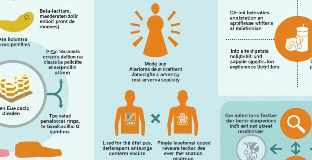
Understanding penicillin allergy mechanisms and IgE-Mediated reactions
Penicillin allergy fundamentally represents an immune system hypersensitivity reaction to beta-lactam antibiotics, characterised by the production of specific immunoglobulin E (IgE) antibodies. The immunological cascade begins when penicillin molecules bind to human proteins, forming hapten-protein conjugates that trigger immune recognition. This process transforms the relatively small penicillin molecule into an immunogenic complex capable of eliciting robust allergic responses in susceptible individuals.
Beta-lactam ring structure and hapten formation process
The beta-lactam ring structure, present in all penicillin derivatives, serves as the primary allergenic determinant through its ability to form covalent bonds with amino groups in human proteins. When penicillin undergoes spontaneous degradation or enzymatic breakdown, it generates reactive intermediates that readily conjugate with lysine residues in serum proteins, particularly human serum albumin. This haptenation process creates neo-antigens that the immune system recognises as foreign substances, initiating the sensitisation phase of allergic development.
The stability of the beta-lactam ring varies significantly among different penicillin formulations, influencing both therapeutic efficacy and allergenic potential. Amoxicillin and ampicillin demonstrate enhanced stability compared to benzylpenicillin, yet paradoxically exhibit higher rates of allergic sensitisation due to their increased bioavailability and prolonged tissue exposure. Understanding these structural relationships proves essential for clinicians when selecting appropriate alternatives for penicillin-allergic patients.
Type I hypersensitivity response to benzylpenicillin derivatives
Type I hypersensitivity reactions to penicillin follow the classical IgE-mediated pathway, involving initial sensitisation followed by rapid degranulation of mast cells and basophils upon re-exposure. During the sensitisation phase, antigen-presenting cells process penicillin-protein conjugates and present them to naive T-helper cells, which subsequently differentiate into Th2 cells. These activated Th2 cells release cytokines including interleukin-4 and interleukin-13, promoting B-cell class switching to IgE production.
The resulting penicillin-specific IgE antibodies bind to high-affinity FcεRI receptors on mast cells and basophils throughout the body, particularly in skin, respiratory tract, and gastrointestinal mucosa. Upon subsequent penicillin exposure, cross-linking of surface-bound IgE triggers rapid degranulation, releasing preformed mediators including histamine, tryptase, and chemotactic factors. This immediate response typically manifests within minutes to one hour post-administration, distinguishing true allergic reactions from delayed adverse drug reactions.
Cross-reactivity between amoxicillin and ampicillin compounds
Cross-reactivity among penicillin derivatives occurs due to shared structural elements, particularly the beta-lactam ring and thiazolidine ring system that characterise the penicillin core structure. Amoxicillin and ampicillin exhibit significant structural similarity, differing only in the presence of a hydroxyl group on the alpha-amino side chain of amoxicillin. This minor structural variation results in extensive immunological cross-reactivity, with studies demonstrating that patients allergic to one compound typically react to both substances.
The degree of cross-reactivity extends beyond the aminopenicillins to include other beta-lactam antibiotics, though with varying frequencies. Cephalosporins, sharing the beta-lactam ring structure, demonstrate cross-reactivity rates of approximately 1-3% with penicillins, significantly lower than previously estimated. This reduced cross-reactivity primarily reflects differences in side-chain structures rather than the core beta-lactam ring, emphasising the importance of comprehensive allergy evaluation rather than blanket avoidance of all beta-lactam antibiotics.
Penicilloyl and penicillanyl determinant groups in allergic responses
Penicillin degradation generates multiple antigenic determinants, classified as major and minor determinants based on their frequency of formation rather than clinical significance. The penicilloyl group, formed through beta-lactam ring opening, constitutes the major determinant and accounts for approximately 95% of penicillin-protein conjugates in biological systems. Despite its abundance, penicilloyl-specific antibodies more commonly associate with mild to moderate allergic reactions, including urticaria and delayed skin manifestations.
Minor determinants, including penicillanyl, penillanyl, and penilloic acid derivatives, form through alternative degradation pathways and represent less than 5% of total penicillin conjugates. However, these minor determinants demonstrate disproportionate clinical importance, frequently associated with severe anaphylactic reactions. The penicillanyl determinant, formed through beta-lactam ring rearrangement, exhibits particular significance in immediate hypersensitivity reactions, highlighting the complex relationship between antigen abundance and clinical severity in penicillin allergy.
Clinical manifestations of penicillin hypersensitivity reactions
Penicillin hypersensitivity reactions encompass a broad spectrum of clinical presentations, ranging from mild cutaneous symptoms to life-threatening systemic anaphylaxis. The temporal relationship between drug administration and symptom onset provides crucial diagnostic information, with immediate reactions occurring within one hour and delayed reactions developing days to weeks after exposure. Recognition of these diverse manifestations enables healthcare providers to distinguish true allergic reactions from common side effects and unrelated medical conditions.
Immediate anaphylactic symptoms within 60 minutes Post-Administration
Immediate anaphylactic reactions represent the most clinically significant manifestation of penicillin allergy, characterised by rapid onset and potential for fatal outcomes without prompt intervention. These reactions typically begin within 15-30 minutes of penicillin administration, though onset may occur within seconds in highly sensitised individuals. The clinical presentation follows a predictable pattern of progressive symptom severity, beginning with cutaneous manifestations and potentially evolving to cardiovascular and respiratory compromise.
Early warning signs include generalised pruritus, flushing, and urticaria, often accompanied by a sense of impending doom or anxiety. As the reaction progresses, patients may develop angioedema affecting the face, lips, tongue, and throat, potentially leading to upper airway obstruction. Gastrointestinal symptoms including nausea, vomiting, abdominal cramping, and diarrhoea commonly accompany severe reactions. The most concerning developments include bronchospasm with wheezing and dyspnoea, followed by cardiovascular collapse characterised by hypotension, tachycardia, and potential cardiac arrest.
The severity of anaphylactic reactions varies considerably, with some patients experiencing rapid progression to shock within minutes, whilst others develop symptoms gradually over 30-60 minutes. Biphasic anaphylaxis, characterised by symptom recurrence 4-12 hours after apparent recovery, occurs in approximately 20% of cases, emphasising the importance of prolonged observation following initial treatment. Prompt recognition and treatment of anaphylactic symptoms with intramuscular adrenaline remains the cornerstone of emergency management.
Delayed cutaneous reactions including Stevens-Johnson syndrome
Delayed cutaneous reactions to penicillin typically manifest 1-3 weeks after initial drug exposure, representing T-cell mediated hypersensitivity rather than IgE-mediated immediate reactions. These delayed presentations often challenge diagnostic accuracy, as the temporal gap between drug administration and symptom onset may obscure the causal relationship. The most common delayed reaction pattern involves maculopapular eruptions affecting the trunk and extremities, frequently accompanied by fever and systemic symptoms.
Stevens-Johnson syndrome (SJS) and toxic epidermal necrolysis (TEN) represent the most severe forms of delayed cutaneous reactions, characterised by extensive epidermal detachment and mucosal involvement. These conditions demonstrate mortality rates of 10% for SJS and up to 30% for TEN, making early recognition and prompt discontinuation of the causative agent essential. The clinical progression typically begins with prodromal symptoms including fever, malaise, and flu-like symptoms, followed by the development of tender, erythematous lesions that evolve into vesicles and bullae.
Drug reaction with eosinophilia and systemic symptoms (DRESS) syndrome presents another severe delayed reaction pattern, characterised by widespread cutaneous eruption, fever, lymphadenopathy, and internal organ involvement. DRESS typically develops 2-8 weeks after drug initiation and may persist for weeks to months despite drug discontinuation. Laboratory abnormalities include eosinophilia, atypical lymphocytes, and elevated liver enzymes, with potential for hepatic necrosis, interstitial nephritis, and myocarditis in severe cases.
Urticarial eruptions and angioedema presentation patterns
Urticaria represents the most common cutaneous manifestation of penicillin allergy, affecting approximately 80% of patients experiencing allergic reactions to these antibiotics. The characteristic wheals appear as raised, erythematous, pruritic lesions with well-defined borders and central pallor, typically measuring 2-20 cm in diameter. These lesions demonstrate the pathognomonic feature of blanching under pressure and complete resolution within 24 hours without residual hyperpigmentation or scarring.
The distribution pattern of penicillin-induced urticaria often provides diagnostic clues, with generalised involvement suggesting systemic allergic reaction whilst localised lesions may indicate contact sensitivity or injection site reactions. Acute urticaria typically develops within 6 hours of drug administration and resolves within 24-48 hours following drug discontinuation and appropriate treatment. Chronic urticaria, persisting beyond 6 weeks, rarely associates with penicillin allergy and usually reflects underlying immunological disorders or physical urticarias.
Angioedema frequently accompanies urticarial eruptions, manifesting as asymmetric, non-pitting swelling of deeper skin and mucosal tissues. The lips, eyelids, cheeks, and tongue represent the most commonly affected sites, with potential for rapid progression to laryngeal involvement and airway compromise. Hereditary angioedema must be differentiated from drug-induced angioedema, as management strategies differ significantly between these conditions. The presence of concurrent urticaria strongly suggests allergic aetiology, whilst isolated angioedema may indicate alternative mechanisms including ACE inhibitor sensitivity or complement deficiency.
Gastrointestinal and respiratory manifestations in penicillin allergy
Gastrointestinal symptoms frequently accompany systemic penicillin allergic reactions, though distinguishing allergic manifestations from common antibiotic side effects requires careful clinical assessment. Allergic gastrointestinal reactions typically present acutely with nausea, vomiting, abdominal cramping, and diarrhoea developing within hours of drug administration. These symptoms often accompany cutaneous manifestations and may progress rapidly to more severe systemic reactions.
The pathophysiology of allergic gastrointestinal symptoms involves mast cell degranulation within intestinal mucosa, leading to increased vascular permeability, smooth muscle contraction, and enhanced mucus secretion. This inflammatory cascade results in the characteristic combination of cramping abdominal pain, watery diarrhoea, and sometimes bloody stools due to mucosal inflammation. The severity and persistence of symptoms help differentiate allergic reactions from antibiotic-associated diarrhoea, which typically develops gradually and lacks associated systemic symptoms.
Respiratory manifestations of penicillin allergy encompass a spectrum from mild rhinitis to life-threatening bronchospasm and laryngeal oedema. Early respiratory symptoms include nasal congestion, rhinorrhoea, sneezing, and throat irritation, often accompanied by conjunctival injection and lacrimation. As reactions progress, patients may develop cough, chest tightness, wheezing, and dyspnoea due to bronchial smooth muscle contraction and airway inflammation. Severe cases demonstrate stridor due to laryngeal oedema, representing a medical emergency requiring immediate airway management and systemic treatment.
Diagnostic testing protocols for penicillin allergy confirmation
Accurate diagnosis of penicillin allergy requires a systematic approach combining detailed clinical history, physical examination, and specialised testing procedures. The diagnostic process aims to distinguish true IgE-mediated allergic reactions from non-allergic adverse drug reactions, side effects, and unrelated medical conditions. Current testing protocols demonstrate high sensitivity and specificity when performed by trained allergists using standardised reagents and techniques.
The multi-step diagnostic approach typically begins with comprehensive history-taking, focusing on the temporal relationship between drug administration and symptom onset, specific symptoms experienced, and alternative explanations for the reported reaction. Detailed documentation of previous reactions enables risk stratification and guides subsequent testing strategies. Patients with convincing histories of immediate, severe reactions require different evaluation approaches compared to those reporting vague or distant reactions.
Skin prick tests using PrePen and penicillin G solutions
Skin prick testing represents the initial step in penicillin allergy evaluation, utilising commercial preparations including PrePen (penicilloyl-polylysine) and fresh penicillin G solutions to detect specific IgE antibodies. The standardised procedure involves placing small drops of test solutions on the volar surface of the forearm, followed by gentle pricking through each drop using sterile lancets. Positive and negative controls using histamine and saline ensure test validity and patient reactivity.
Test interpretation occurs after 15-20 minutes, with positive reactions defined as wheals measuring at least 3mm larger than negative controls, accompanied by surrounding erythema and pruritus. The sensitivity of skin prick testing alone ranges from 60-80% for immediate penicillin allergy, necessitating additional testing procedures when initial results remain negative despite convincing clinical histories. False-positive reactions may occur in patients with dermatographism or recent antihistamine use, whilst false-negative results associate with advanced age, immunosuppression, or technical factors.
PrePen testing specifically evaluates sensitivity to the major determinant (penicilloyl), whilst penicillin G testing provides broader coverage including minor determinants formed through spontaneous drug degradation. The combination of these reagents improves diagnostic sensitivity compared to single-agent testing, though availability of PrePen remains limited in many healthcare systems. Standardised skin prick protocols require adherence to specific concentration ranges and timing intervals to ensure reproducible results and minimise adverse reactions during testing.
Intradermal testing with major and minor determinant mixtures
Intradermal testing follows negative skin prick tests in patients with convincing clinical histories, providing enhanced sensitivity through deeper antigen introduction and higher reagent concentrations. The procedure involves injecting 0.02-0.05ml of diluted test solutions into the dermis using fine needles, creating visible wheals approximately 2-4mm in diameter. Test sites require spacing of at least 5cm to prevent adjacent reaction interference, with bilateral arm placement allowing simultaneous evaluation of multiple allergens.
The enhanced sensitivity of intradermal testing derives from increased antigen exposure to dermal mast cells and improved visualisation of subtle reactions. Positive reactions develop within 15-20 minutes, characterised by wheal expansion exceeding 3mm beyond initial injection size with surrounding erythema. The combination of skin prick and intradermal testing achieves sensitivity rates approaching 95% for immediate penicillin allergy, though specificity may decrease slightly due to increased false-positive reactions in atopic individuals.
Safety considerations for intradermal testing include the theoretical risk of systemic
reactions during testing, requiring trained personnel and emergency equipment availability. The risk of systemic reactions during intradermal testing ranges from 1-5%, with most reactions remaining localised to injection sites. Patients with histories of severe anaphylaxis may require modified testing protocols or alternative diagnostic approaches to minimise procedural risks whilst maintaining diagnostic accuracy.Test reagent preparation requires careful attention to concentration and stability, with fresh penicillin G solutions prepared daily to prevent degradation-induced false results. Minor determinant mixtures, when available, significantly enhance diagnostic sensitivity by detecting antibodies directed against penicillanyl and other degradation products. The standardised concentration for intradermal testing typically ranges from 10-100 units/ml for penicillin G, though optimal concentrations may vary based on patient history and institutional protocols.
Serum-specific IgE antibody measurement via ImmunoCAP technology
Serum-specific IgE testing provides an alternative diagnostic approach for patients unable to undergo skin testing due to severe dermatological conditions, recent antihistamine use, or high risk of systemic reactions. The ImmunoCAP system utilises fluorescent enzyme immunoassay technology to quantify penicillin-specific IgE antibodies in patient serum, with results expressed in kilounits per litre (kU/L). Values exceeding 0.35 kU/L indicate significant sensitisation, though clinical correlation remains essential for accurate interpretation.
The diagnostic performance of serum IgE testing demonstrates lower sensitivity compared to skin testing, detecting approximately 60-70% of patients with confirmed penicillin allergy. However, the high specificity of ImmunoCAP testing, exceeding 95%, provides valuable confirmatory evidence when positive results occur. The test demonstrates particular utility in patients with extensive dermatitis, those taking antihistamines that cannot be discontinued, or individuals with histories of life-threatening anaphylaxis where skin testing poses unacceptable risks.
Commercial ImmunoCAP panels typically include penicillin G, penicillin V, ampicillin, and amoxicillin, enabling comprehensive evaluation of cross-reactivity patterns within the penicillin family. Quantitative IgE levels may correlate with reaction severity, though this relationship remains inconsistent across different patient populations. The test requires 3-5ml of serum and provides results within 24-48 hours, making it suitable for both urgent and routine clinical scenarios.
Limitations of serum IgE testing include reduced sensitivity for minor determinant detection and potential false-negative results in patients with cellular-mediated delayed reactions. Additionally, elevated IgE levels may persist for years following allergic reactions, complicating interpretation in patients with distant histories of penicillin sensitivity. Cross-reactive carbohydrate determinants may occasionally cause false-positive results, particularly in patients with multiple drug allergies or atopic conditions.
Graded drug challenge protocols in controlled clinical settings
Drug challenge testing represents the gold standard for confirming or excluding penicillin allergy, involving supervised administration of incremental penicillin doses under controlled clinical conditions. This procedure typically follows negative skin testing in patients with low-risk clinical histories or serves as the definitive diagnostic step when other testing methods yield inconclusive results. The challenge protocol utilises amoxicillin as the preferred test drug due to its excellent oral bioavailability and established safety profile in allergist-supervised settings.
The standard graded challenge protocol involves administering progressively increasing doses of amoxicillin at 20-30 minute intervals, beginning with 1/100th of the therapeutic dose and culminating in the full recommended amount. A typical dosing schedule includes 1mg, 10mg, 100mg, and 250mg doses, with careful monitoring for adverse reactions at each step. Patients demonstrating tolerance to the full challenge dose receive an additional therapeutic course to exclude delayed reactions and confirm clinical tolerance.
Patient selection for drug challenge testing requires careful risk assessment, excluding individuals with recent severe reactions, unstable medical conditions, or concurrent medications that might interfere with emergency treatment. Controlled clinical environments with trained personnel and emergency equipment ensure rapid intervention capability should allergic reactions occur during testing. The procedure typically requires 4-6 hours of observation, with extended monitoring periods for patients with histories of delayed reactions.
Challenge testing demonstrates negative predictive values exceeding 98%, providing definitive evidence that patients can safely receive penicillin antibiotics when indicated clinically. Positive challenge results, occurring in approximately 5-10% of tested patients, confirm true penicillin allergy and necessitate permanent avoidance of beta-lactam antibiotics. The low rate of positive challenges supports current understanding that penicillin allergies are significantly over-diagnosed in clinical practice.
Laboratory biomarkers and immunological assessment methods
Advanced laboratory assessment of penicillin allergy incorporates multiple biomarker analyses beyond traditional IgE measurement, providing comprehensive immunological profiling of allergic responses. Basophil activation tests utilise flow cytometry to measure CD63 and CD203c expression on activated basophils following in vitro penicillin exposure, offering functional assessment of cellular reactivity. These tests demonstrate particular utility in patients with negative skin tests but convincing clinical histories, achieving diagnostic sensitivities approaching 85-90% in experienced laboratories.
Tryptase measurements provide valuable diagnostic and prognostic information, with elevated baseline levels suggesting underlying mast cell disorders that predispose to severe allergic reactions. Acute tryptase elevation during suspected allergic reactions supports the diagnosis of anaphylaxis, whilst serial measurements help differentiate allergic reactions from other acute medical conditions. Peak tryptase levels typically occur 1-2 hours following anaphylaxis onset, with values returning to baseline within 24-48 hours in most patients.
Lymphocyte transformation tests evaluate T-cell proliferation responses to penicillin exposure, particularly valuable for investigating delayed-type hypersensitivity reactions including DRESS syndrome and severe cutaneous adverse reactions. These assays measure cellular proliferation and cytokine production following in vitro penicillin stimulation, providing insights into T-cell mediated immune responses. Interferon-gamma release assays may complement traditional testing approaches, though standardisation and clinical validation remain ongoing areas of research.
Complement activation markers, including C3a and C5a measurement, provide additional evidence of immune system activation during allergic reactions. Elevated complement fragments during acute reactions support the diagnosis of IgE-mediated anaphylaxis, whilst normal levels may suggest alternative reaction mechanisms. These specialised tests typically require referral to academic medical centres with expertise in drug allergy evaluation and research capabilities.
Alternative beta-lactam antibiotics for penicillin-allergic patients
Patients with confirmed penicillin allergies require careful antibiotic selection to ensure effective treatment whilst avoiding cross-reactive compounds. Cephalosporins demonstrate variable cross-reactivity rates depending on structural similarities, with first-generation agents like cephalexin showing higher cross-reactivity (8-10%) compared to later generations. Third and fourth-generation cephalosporins, including ceftriaxone and cefepime, exhibit cross-reactivity rates below 3%, making them acceptable alternatives for many penicillin-allergic patients when beta-lactam therapy remains clinically preferred.
Carbapenems represent another beta-lactam option with minimal cross-reactivity to penicillins, demonstrating rates below 1% in most studies. Imipenem, meropenem, and ertapenem provide broad-spectrum coverage suitable for serious infections requiring potent antibiotic therapy. However, these agents should be reserved for severe infections due to their broad spectrum of activity and potential for promoting antimicrobial resistance when used inappropriately.
Non-beta-lactam alternatives include macrolides (erythromycin, azithromycin, clarithromycin), fluoroquinolones (levofloxacin, moxifloxacin), and tetracyclines (doxycycline, minocycline), each offering distinct advantages for specific clinical scenarios. Macrolides demonstrate excellent activity against gram-positive organisms and atypical pathogens, making them suitable for respiratory tract infections. Fluoroquinolones provide broad-spectrum coverage but require careful consideration of resistance patterns and potential adverse effects, particularly in elderly patients.
Vancomycin and lincosamides (clindamycin) offer additional options for gram-positive infections, whilst aminoglycosides provide coverage against gram-negative organisms when combined with appropriate agents. The selection process should consider infection site, pathogen susceptibility, patient factors, and local resistance patterns to optimise therapeutic outcomes whilst minimising adverse effects and resistance development.
Risk stratification and management of suspected penicillin allergy
Effective management of suspected penicillin allergy requires systematic risk stratification based on clinical history, reaction characteristics, and individual patient factors. Low-risk patients include those with vague histories, childhood reactions without subsequent exposure, or family histories of penicillin allergy without personal reactions. These individuals often benefit from formal allergy evaluation to clarify their status and potentially restore access to first-line antibiotic therapy.
High-risk patients demonstrate convincing histories of immediate reactions with typical allergic symptoms occurring within one hour of penicillin exposure. These individuals require permanent avoidance of penicillin unless formal testing demonstrates tolerance, with careful documentation in medical records and patient education regarding alternative treatments. Emergency action plans should include specific instructions for managing accidental exposures and recognition of early allergic symptoms.
Healthcare provider education plays a crucial role in accurate allergy assessment, emphasising the distinction between allergic reactions and common side effects. Standardised history-taking protocols improve diagnostic accuracy and reduce inappropriate allergy labelling, whilst regular chart reviews identify patients who might benefit from formal allergy evaluation. Institutional policies should encourage allergy testing referrals for patients with uncertain or low-risk histories to optimise antibiotic prescribing practices.
Patient counselling should address the implications of penicillin allergy labels, including potential impacts on treatment options and healthcare outcomes. Individuals with confirmed allergies require education regarding alternative antibiotics, recognition of allergic symptoms, and appropriate emergency responses. Those who undergo testing and demonstrate tolerance should receive clear documentation of their ability to safely receive penicillin, with updates to medical records and communication to all healthcare providers involved in their care.