The persistent need to urinate frequently whilst only producing small volumes of urine represents one of the most common and distressing urological complaints encountered in clinical practice. This condition, medically termed pollakiuria, affects millions of individuals worldwide and can significantly impact quality of life, sleep patterns, and daily activities. Unlike normal voiding patterns where the average person produces 200-500ml per void, those experiencing this condition may find themselves visiting the toilet every 30-60 minutes, often producing less than 50-100ml of urine each time.
Understanding the underlying mechanisms behind frequent small-volume urination requires a comprehensive examination of bladder physiology, neurological control systems, and the various pathological processes that can disrupt normal voiding function. The complexity of this symptom stems from its multifactorial nature, involving everything from simple infections to complex neurological disorders, hormonal imbalances, and structural abnormalities of the urogenital system.
Medical terminology: understanding pollakiuria and urinary frequency disorders
Defining pollakiuria: clinical classification of frequent Small-Volume urination
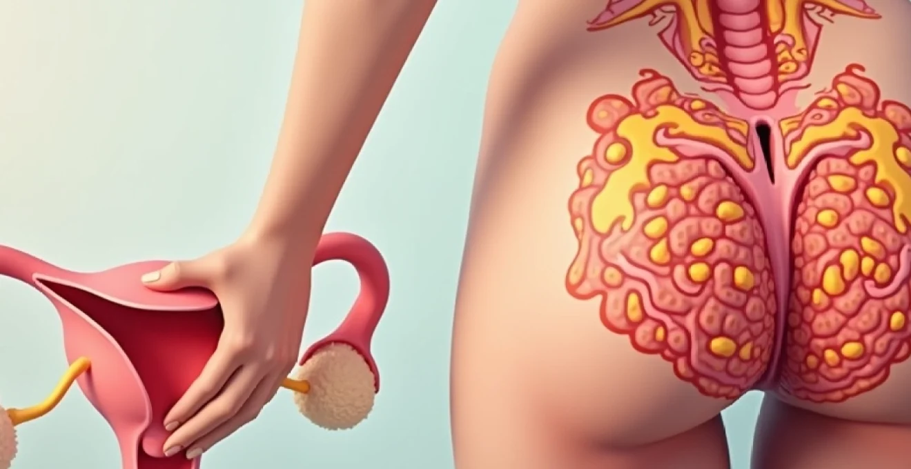
Pollakiuria, derived from the Greek words “pollakis” meaning often and “ouron” meaning urine, represents the medical term for abnormally frequent urination characterised by small volumes. This condition differs significantly from polyuria, where patients produce large volumes of urine, often exceeding 3 litres per day. In pollakiuria, the total daily urine output typically remains within normal limits (1.5-2 litres), but the frequency of voiding episodes increases dramatically, sometimes reaching 20-30 times per day.
The clinical significance of pollakiuria extends beyond mere inconvenience, as it often indicates underlying pathology affecting the bladder’s storage function. Healthcare professionals classify this condition based on several parameters, including void frequency, individual void volumes, presence of associated symptoms such as urgency or dysuria, and the impact on nocturnal sleep patterns. Understanding these classifications helps clinicians develop targeted diagnostic strategies and appropriate treatment protocols.
Distinguishing between urinary frequency, urgency, and nocturia symptoms
While often occurring together, urinary frequency, urgency, and nocturia represent distinct clinical entities with specific diagnostic criteria. Urinary frequency refers to voiding more than 8 times during waking hours, regardless of the urgency level or volume produced. This symptom can occur without any accompanying sensations of urgency, particularly in cases involving bladder outlet obstruction or incomplete emptying.
Urgency, conversely, describes the sudden, compelling desire to urinate that proves difficult to defer. This sensation often accompanies pollakiuria in conditions such as overactive bladder syndrome or acute cystitis. Nocturia specifically refers to awakening from sleep one or more times to urinate, with normal sleep subsequently resumed. The distinction between these symptoms proves crucial for accurate diagnosis, as different underlying pathologies may present with varying combinations of these complaints.
Normal bladder capacity parameters and voiding patterns in adults
Adult bladder capacity typically ranges between 400-600ml, with the first urge to void occurring at approximately 200-300ml of bladder filling. Normal voiding patterns involve 6-8 urinations per 24-hour period, with individual void volumes averaging 250-400ml. The bladder’s remarkable ability to accommodate increasing volumes whilst maintaining low intravesical pressures depends on the coordinated function of detrusor muscle fibres and intact neurological pathways.
Deviations from these normal parameters often indicate pathological processes. When void volumes consistently fall below 150ml despite normal fluid intake, this suggests either reduced functional bladder capacity or incomplete emptying with frequent overflow. These measurements serve as crucial baseline data for clinicians evaluating patients with suspected pollakiuria, helping differentiate between various underlying causes and guiding appropriate diagnostic investigations.
Post-void residual volume measurements and clinical significance
Post-void residual (PVR) volume represents the amount of urine remaining in the bladder immediately after voluntary urination. Normal PVR values should remain below 50ml in healthy adults, though values up to 100ml may be acceptable in elderly patients. Elevated PVR measurements often accompany pollakiuria, particularly in cases involving bladder outlet obstruction or detrusor underactivity.
Measuring PVR accurately requires either bladder ultrasonography or catheterisation immediately following voiding. This parameter proves invaluable in distinguishing between storage disorders (low PVR) and emptying disorders (high PVR) in patients presenting with frequent small-volume urination. Understanding PVR significance enables healthcare providers to tailor treatment strategies appropriately, addressing either storage dysfunction or emptying impairment as the primary therapeutic target.
Pathophysiological mechanisms behind reduced bladder capacity and incomplete emptying
Detrusor muscle dysfunction and overactive bladder syndrome pathways
Detrusor overactivity represents one of the most common pathophysiological mechanisms underlying pollakiuria, characterised by involuntary detrusor contractions during the filling phase of the micturition cycle. These abnormal contractions create urgency sensations and reduce functional bladder capacity, forcing patients to void small volumes frequently to prevent incontinence episodes. The underlying mechanisms involve alterations in muscarinic receptor sensitivity, changes in intracellular calcium handling, and modifications in smooth muscle cell coupling.
Overactive bladder syndrome affects approximately 17% of adults, with prevalence increasing significantly with age. The condition involves complex interactions between peripheral bladder mechanisms and central nervous system processing of afferent signals. Research suggests that abnormal urothelial signalling plays a crucial role, with increased release of mediators such as adenosine triphosphate (ATP) and acetylcholine contributing to heightened detrusor sensitivity and spontaneous contractions.
Neurogenic bladder conditions: multiple sclerosis and spinal cord injury effects
Neurological disorders frequently disrupt the delicate balance between bladder storage and emptying functions, often resulting in pollakiuria accompanied by urgency and incomplete evacuation. Multiple sclerosis affects bladder function in approximately 80% of patients, typically presenting with detrusor overactivity, detrusor-sphincter dyssynergia, or combinations thereof. The demyelinating plaques characteristic of MS can affect any portion of the neuraxis controlling micturition, from the pontine micturition centre to peripheral nerve pathways.
Spinal cord injuries produce varying patterns of bladder dysfunction depending on the level and completeness of the lesion. Suprasacral injuries typically result in detrusor overactivity with detrusor-sphincter dyssynergia, creating a clinical picture of frequent small voids accompanied by high post-void residuals. The neuroplasticity following spinal cord injury can lead to progressive changes in bladder function over time, necessitating ongoing monitoring and treatment adjustments to prevent complications such as autonomic dysreflexia or upper tract deterioration.
Bladder outlet obstruction: benign prostatic hyperplasia and urethral strictures
Bladder outlet obstruction creates a complex pathophysiological cascade that frequently manifests as pollakiuria, particularly in men with benign prostatic hyperplasia (BPH). The obstructed bladder initially compensates through detrusor hypertrophy and increased contractile force, but chronic obstruction eventually leads to detrusor decompensation, reduced compliance, and the development of secondary detrusor overactivity. This progression explains why men with BPH often experience both obstructive symptoms (weak stream, hesitancy) and irritative symptoms (frequency, urgency, nocturia).
Urethral strictures, whether congenital, inflammatory, or traumatic in origin, create similar obstructive patterns but may present at any age and in both sexes. The degree of outlet obstruction correlates with the severity of compensatory changes, including bladder wall thickening, trabeculation formation, and eventually diverticulum development. Understanding these progressive changes helps clinicians appreciate why early intervention often proves more effective than delayed treatment in preserving long-term bladder function.
Inflammatory processes: interstitial cystitis and chronic pelvic pain syndrome
Interstitial cystitis/bladder pain syndrome (IC/BPS) represents a chronic inflammatory condition characterised by bladder pain, pressure, and discomfort associated with urinary frequency and urgency. The pathophysiology involves urothelial dysfunction, mast cell activation, neurogenic inflammation, and potentially autoimmune mechanisms. Patients with IC/BPS typically void small volumes (50-150ml) frequently throughout the day and night, as bladder filling beyond minimal volumes produces significant discomfort.
The inflammatory cascade in IC/BPS involves increased vascular permeability, mast cell degranulation, and release of inflammatory mediators including histamine, prostaglandins, and neurotrophins. These substances sensitise afferent nerve pathways, creating a cycle of pain, frequency, and further inflammation. Chronic pelvic pain syndrome in men presents similar symptoms but may involve prostatitis, pelvic floor dysfunction, or referred pain from other pelvic structures, making accurate diagnosis challenging without comprehensive evaluation.
Infectious and inflammatory causes of frequent Small-Volume urination
Acute cystitis: escherichia coli and staphylococcus saprophyticus infections
Acute bacterial cystitis remains the most common cause of sudden-onset pollakiuria, particularly affecting women of reproductive age. Escherichia coli accounts for approximately 85% of uncomplicated urinary tract infections, utilising specific adhesins to bind to uroepithelial cells and establish infection despite normal voiding mechanisms. The inflammatory response triggered by bacterial invasion creates mucosal oedema, increased vascular permeability, and heightened sensory nerve activity, resulting in the classic triad of frequency, urgency, and dysuria.
Staphylococcus saprophyticus represents the second most common uropathogen in young, sexually active women, particularly during summer months and following sexual activity. Unlike E. coli infections, S. saprophyticus demonstrates unique tropism for the urogenital tract and may present with more subtle symptoms initially. The bacterial virulence factors contribute to mucosal invasion and inflammatory cascade initiation, creating the characteristic clinical presentation of frequent, painful urination with small void volumes rarely exceeding 100ml during acute phases.
Chronic urethritis: chlamydia trachomatis and mycoplasma genitalium
Chronic urethritis caused by sexually transmitted pathogens often presents with persistent pollakiuria that may develop gradually over weeks to months. Chlamydia trachomatis, the most common bacterial sexually transmitted infection globally, demonstrates particular affinity for columnar epithelium lining the urethra and can establish persistent, low-grade inflammation even in the absence of overt symptoms. The chronic inflammatory process affects urethral sensation and may create reactive changes in the bladder neck and trigone, contributing to frequency symptoms.
Mycoplasma genitalium, increasingly recognised as a significant uropathogen, produces chronic urethritis characterised by minimal discharge but persistent irritative voiding symptoms. The organism’s unique cell wall structure makes it inherently resistant to many conventional antibiotics, often resulting in treatment failures and chronic symptoms. These atypical pathogens require specific diagnostic approaches, including nucleic acid amplification testing, as they frequently remain undetected by routine urine cultures, leading to delayed diagnosis and inappropriate antibiotic therapy.
Prostatitis syndromes: bacterial and chronic pelvic pain classifications
Prostatitis syndromes encompass a heterogeneous group of conditions affecting men of all ages, with chronic prostatitis/chronic pelvic pain syndrome (CP/CPPS) being the most prevalent form. Acute bacterial prostatitis, though less common, presents with severe systemic symptoms accompanied by marked pollakiuria, as the inflamed prostate gland compresses the proximal urethra and creates secondary bladder outlet obstruction. The condition requires immediate antibiotic therapy to prevent complications such as abscess formation or sepsis.
Chronic bacterial prostatitis involves recurrent episodes of bacterial infection within the prostate gland, often presenting with persistent lower urinary tract symptoms including frequency, urgency, and pelvic discomfort. The most challenging form, CP/CPPS, affects approximately 10% of men at some point in their lives and presents with chronic pelvic pain accompanied by voiding dysfunction in the absence of demonstrable bacterial infection. The complex pathophysiology of CP/CPPS likely involves neuromuscular dysfunction, immune system dysregulation, and possibly infectious agents that remain undetectable by current diagnostic methods.
Sexually transmitted infections: gonorrhoea and trichomonas vaginalis impact
Neisseria gonorrhoeae infection typically presents with acute urethritis characterised by purulent discharge and severe dysuria, though some patients may experience primarily frequency symptoms with minimal discharge. The gram-negative diplococcus demonstrates remarkable ability to evade host immune responses through antigenic variation and can rapidly ascend the urogenital tract, potentially causing epididymitis in men or pelvic inflammatory disease in women. The acute inflammatory response creates significant urethral oedema and hypersensitivity, resulting in frequent small-volume voids often accompanied by haematuria.
Trichomonas vaginalis, a flagellated protozoan parasite, affects both men and women but often remains asymptomatic in male partners. In women, the infection typically involves the vagina, urethra, and periurethral glands, creating a clinical presentation that may include pollakiuria, urgency, and dysuria alongside the characteristic frothy, malodorous vaginal discharge. The parasitic infection can persist for months or years if untreated, creating chronic inflammatory changes that may contribute to recurrent urinary tract infections and persistent lower urinary tract symptoms requiring comprehensive evaluation and partner treatment.
Hormonal and metabolic disorders contributing to urinary frequency
Diabetes mellitus represents one of the most significant metabolic disorders contributing to urinary frequency, though the mechanism differs from typical pollakiuria. In uncontrolled diabetes, hyperglycaemia leads to osmotic diuresis, where excess glucose in the urine draws additional water, resulting in polyuria rather than true pollakiuria. However, diabetic patients frequently develop secondary complications including diabetic cystopathy, characterised by impaired bladder sensation, increased bladder capacity, and incomplete emptying that can present with overflow-type frequency symptoms.
Hormonal fluctuations, particularly oestrogen deficiency in postmenopausal women, significantly impact urogenital tissues and contribute to frequency symptoms. Oestrogen receptors throughout the lower urinary tract become less stimulated, leading to urethral atrophy, decreased urethral closure pressure, and altered bladder sensitivity. These changes often manifest as a combination of stress incontinence and irritative voiding symptoms, including increased frequency and urgency. The complex interplay between hormonal status and bladder function explains why hormone replacement therapy may provide symptom relief in carefully selected postmenopausal women experiencing bothersome lower urinary tract symptoms.
Hyperthyroidism can contribute to urinary frequency through multiple mechanisms, including increased metabolic rate, enhanced cardiac output, and direct effects on bladder smooth muscle contractility. Thyroid hormones influence beta-adrenergic receptor sensitivity, potentially affecting the normal inhibitory sympathetic input to the detrusor muscle during the storage phase. Additionally, hyperthyroid patients often experience increased fluid intake due to heat intolerance and increased metabolic demands, creating a secondary contributor to frequency symptoms that resolves with appropriate thyroid function normalisation.
Diagnostic evaluation protocols for persistent Small-Volume urination
Comprehensive evaluation of patients presenting with persistent small-volume urination begins with detailed history-taking that encompasses symptom onset, progression patterns, associated symptoms, fluid intake habits, medication review, and impact on quality of life. Bladder diaries prove invaluable in quantifying symptoms objectively, recording voided volumes, timing of voids, fluid intake, and associated symptoms over 3-7 consecutive days. These diaries often reveal patterns invisible to patients, such as nocturnal polyuria, excessive caffeine intake, or correlation between symptoms and specific activities or stressors.
Physical examination should include abdominal palpation for bladder distension or masses, pelvic examination in women to assess for prolapse or urogenital atrophy, digital rectal examination in men to evaluate prostate
size, consistency, and nodularity, and focused neurological assessment when neurogenic causes are suspected. The combination of history, physical examination, and initial diagnostic studies helps establish the differential diagnosis and guide subsequent testing protocols.
Laboratory evaluation typically commences with comprehensive urinalysis and urine culture, which remain fundamental in excluding infectious aetiologies. Microscopic examination may reveal pyuria, bacteriuria, or crystals that provide diagnostic clues, whilst nitrite and leucocyte esterase testing offers rapid screening for bacterial infection. Post-void residual measurement via bladder ultrasonography provides crucial information about emptying efficiency and helps differentiate between storage and voiding dysfunction. Advanced diagnostic modalities such as urodynamic studies, cystoscopy, or imaging studies may be indicated based on initial findings and clinical suspicion of specific underlying pathologies.
Urodynamic testing provides objective assessment of bladder and urethral function, measuring parameters including detrusor pressure, bladder compliance, maximum flow rate, and detrusor-sphincter coordination. These studies prove particularly valuable when conservative treatments fail or when neurological causes are suspected. Cystoscopy allows direct visualisation of the bladder mucosa and urethra, enabling detection of inflammatory changes, stones, tumours, or structural abnormalities that may contribute to frequency symptoms. The timing and selection of these invasive procedures require careful consideration of patient symptoms, quality of life impact, and likelihood of discovering treatable pathology.
Evidence-based treatment approaches and management strategies
Management of persistent small-volume urination follows a stepwise approach, beginning with conservative interventions and progressing to more invasive therapies based on symptom severity, underlying aetiology, and treatment response. Behavioural modifications form the cornerstone of initial management, including scheduled voiding regimens, bladder training programmes, and pelvic floor muscle exercises. Fluid management strategies involve optimising total intake whilst avoiding excessive consumption, particularly of bladder irritants such as caffeine, alcohol, and acidic beverages.
Pharmacological interventions target specific pathophysiological mechanisms underlying the patient’s symptoms. Antimuscarinic agents, including oxybutynin, tolterodine, and solifenacin, effectively reduce detrusor overactivity and improve storage symptoms in patients with overactive bladder syndrome. Beta-3 adrenergic agonists such as mirabegron offer alternative mechanisms of action with potentially fewer anticholinergic side effects. Antibiotic therapy remains essential for confirmed bacterial infections, with selection based on local resistance patterns and patient-specific factors such as allergy history and renal function.
Advanced therapeutic options include intravesical treatments, neuromodulation techniques, and surgical interventions reserved for refractory cases. Botulinum toxin injections into the detrusor muscle provide effective symptom relief for patients with overactive bladder unresponsive to oral medications, typically lasting 6-9 months per treatment cycle. Sacral neuromodulation offers another option for carefully selected patients, involving implantation of electrodes that modulate neural pathways controlling bladder function. These interventions require comprehensive patient counselling regarding risks, benefits, and realistic expectations for symptom improvement.
The management approach must be individualised based on patient age, comorbidities, symptom severity, and personal preferences. Regular follow-up assessments help monitor treatment response and identify the need for therapy modifications or advancement to more invasive options. Quality of life measures and objective parameters such as voiding diary data guide treatment decisions and help optimise outcomes. Multidisciplinary collaboration involving urologists, primary care physicians, and sometimes gynaecologists or neurologists ensures comprehensive care addressing all contributing factors to the patient’s symptom complex.
Patient education plays a crucial role in successful management, helping individuals understand their condition, treatment options, and realistic expectations for improvement. Many patients benefit from lifestyle modifications that extend beyond fluid management, including weight reduction, smoking cessation, and management of contributing medical conditions such as diabetes or constipation. The chronic nature of many conditions causing pollakiuria necessitates long-term management strategies that balance symptom control with treatment burden and potential adverse effects.