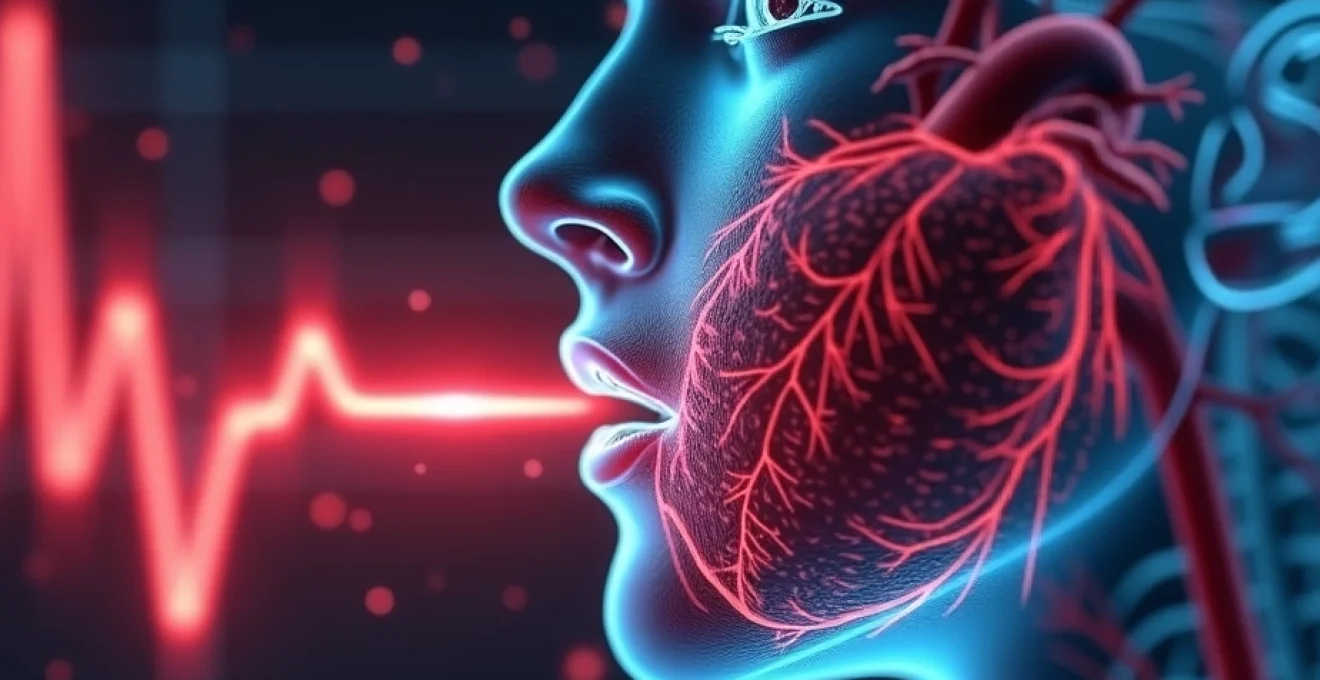
Every time you take a breath, your heart responds in a remarkably sophisticated dance of physiological coordination. This natural phenomenon, where your heart rate increases during inspiration and decreases during expiration, represents one of the most elegant examples of how your cardiovascular and respiratory systems work together. Far from being a medical anomaly, this rhythmic variation in heart rate demonstrates the healthy functioning of your autonomic nervous system and reflects your body’s remarkable ability to optimise oxygen delivery throughout each breathing cycle.
The mechanism behind this heart rate variation involves complex interactions between your nervous system, blood vessels, and cardiac chambers. Understanding why your heart accelerates when you inhale provides fascinating insights into human physiology and reveals how your body constantly fine-tunes its performance to meet metabolic demands. This respiratory-cardiac coupling occurs in healthy individuals of all ages, though it’s particularly pronounced in young, fit individuals with robust autonomic nervous system function.
Respiratory sinus arrhythmia: the physiological mechanism behind heart rate variability
Respiratory sinus arrhythmia represents the normal, healthy variation in heart rate that occurs with breathing. This phenomenon demonstrates the intricate relationship between your respiratory and cardiovascular systems, where each inhalation triggers a predictable acceleration of your heart rate. The term “arrhythmia” might sound concerning, but in this context, it simply describes the rhythmic irregularity that occurs naturally with breathing cycles.
The magnitude of this heart rate variation typically ranges from 5 to 20 beats per minute in healthy adults, with younger individuals often displaying more pronounced variations. This variability reflects the health and responsiveness of your autonomic nervous system, which controls involuntary bodily functions including heart rate, breathing, and digestion. Research indicates that reduced heart rate variability can be an early indicator of cardiovascular disease , making respiratory sinus arrhythmia an important marker of cardiac health.
Parasympathetic nervous system modulation during inspiratory phases
During inspiration, your parasympathetic nervous system undergoes temporary suppression, leading to the characteristic increase in heart rate. The parasympathetic system, often called the “rest and digest” division of your autonomic nervous system, normally acts as a brake on your heart rate through the release of acetylcholine at the sinoatrial node. When you inhale, this braking effect diminishes, allowing your heart rate to accelerate naturally.
This modulation occurs through the vagus nerve, the longest cranial nerve that connects your brain to various organs including your heart. The vagal influence on heart rate follows a predictable pattern throughout your breathing cycle, with maximum suppression occurring during peak inspiration . This precise timing ensures that your heart can pump more blood when your lungs are filling with oxygen-rich air, optimising the efficiency of oxygen transport throughout your body.
Vagal tone suppression and cardiac acceleration patterns
Vagal tone refers to the baseline level of parasympathetic activity affecting your heart rate. During inspiration, this tone undergoes systematic suppression, creating the characteristic acceleration pattern observed in respiratory sinus arrhythmia. The degree of vagal tone suppression directly correlates with the magnitude of heart rate increase, explaining why individuals with higher baseline vagal tone often display more pronounced respiratory sinus arrhythmia.
The temporal relationship between breathing and heart rate acceleration follows a precise pattern. Heart rate begins to increase approximately 200-300 milliseconds after the onset of inspiration, reaching peak acceleration during mid-inspiration. This timing allows your cardiovascular system to anticipate and respond to the increased venous return that occurs during the inspiratory phase, demonstrating the remarkable coordination between your respiratory and circulatory systems.
Baroreceptor reflex integration with respiratory cycles
Baroreceptors, specialised pressure sensors located in your carotid arteries and aortic arch, play a crucial role in modulating the respiratory sinus arrhythmia response. These receptors continuously monitor blood pressure and send signals to your brainstem to adjust heart rate accordingly. During inspiration, changes in intrathoracic pressure affect the sensitivity and response of these baroreceptors, contributing to the overall heart rate variability pattern.
The integration of baroreceptor feedback with respiratory cycles creates a sophisticated control mechanism that helps maintain optimal blood pressure throughout breathing. When you inhale deeply, the temporary reduction in baroreceptor sensitivity allows for the heart rate increase necessary to accommodate increased venous return. This coordinated response ensures that blood pressure remains stable despite the mechanical effects of breathing on your cardiovascular system .
Sinoatrial node response to autonomic neural fluctuations
The sinoatrial node, your heart’s natural pacemaker, serves as the final common pathway for autonomic influences on heart rate. This specialised cluster of cells responds rapidly to changes in sympathetic and parasympathetic input, translating neural signals into the rhythmic electrical impulses that drive your heartbeat. During inspiration, the sinoatrial node experiences reduced parasympathetic inhibition, allowing its intrinsic firing rate to increase.
The cellular mechanisms underlying this response involve complex interactions between ion channels and neurotransmitter receptors within the sinoatrial node. Acetylcholine released by parasympathetic nerve terminals normally slows the node’s firing rate by affecting potassium and calcium channels. When parasympathetic activity decreases during inspiration, these channels return towards their baseline state, naturally accelerating the pacemaker’s rhythm and increasing your heart rate.
Cardiopulmonary coupling: venous return dynamics during inspiration
The mechanical act of breathing creates profound effects on blood flow throughout your cardiovascular system, with inspiration generating a powerful suction effect that enhances venous return to your heart. This phenomenon, known as the respiratory pump, occurs because the pressure changes within your thoracic cavity during breathing directly influence blood flow in the great veins returning to your heart. When you inhale, the expansion of your chest cavity creates negative pressure that literally draws blood from your peripheral circulation towards your heart.
This enhanced venous return during inspiration represents one of the primary reasons why your heart rate must increase during this phase of breathing. The additional blood volume returning to your heart requires increased cardiac output to maintain circulatory efficiency . Your cardiovascular system has evolved this elegant solution of coupling heart rate increases with inspiratory venous return enhancement, ensuring that the extra blood entering your heart is promptly circulated throughout your body.
Thoracic pressure changes and right ventricular preload enhancement
During inspiration, your diaphragm descends and your ribcage expands, creating negative pressure within your thoracic cavity. This pressure reduction has immediate effects on the great veins entering your heart, particularly the superior and inferior vena cavae. The pressure gradient between your peripheral circulation and the thoracic cavity increases significantly, accelerating blood flow towards your right ventricle and increasing what physiologists term “preload” – the volume of blood filling your heart before it contracts.
The magnitude of this preload enhancement can increase right ventricular filling by 20-30% during deep inspiration compared to expiration. This substantial increase in blood volume requires your heart to work harder and faster to maintain effective circulation. The physiological response involves not only an increase in heart rate but also adjustments in the force of cardiac contraction to handle the increased blood volume efficiently.
Frank-starling mechanism activation through increased venous return
The Frank-Starling mechanism represents a fundamental property of cardiac muscle whereby increased filling of the heart leads to more forceful contractions. During inspiration, the enhanced venous return triggers this mechanism, causing your heart to contract more vigorously to eject the additional blood volume. However, relying solely on increased contractile force would be metabolically expensive and potentially insufficient to handle the volume load effectively.
The concurrent increase in heart rate during inspiration works synergistically with the Frank-Starling mechanism to optimise cardiac output. By increasing both the force and frequency of contractions, your heart can handle the inspiratory surge in venous return without compromising circulatory efficiency. This dual response ensures that oxygen delivery to your tissues remains optimal throughout all phases of the breathing cycle .
Interventricular septal shift effects on left ventricular filling
The increased filling of your right ventricle during inspiration doesn’t occur in isolation but affects your left ventricle through mechanical interactions within the shared pericardial space. As your right ventricle fills with additional blood during inspiration, the interventricular septum shifts slightly towards the left ventricle, temporarily reducing left ventricular filling capacity. This phenomenon, known as ventricular interdependence, creates complex dynamics in cardiac function during breathing.
Paradoxically, this temporary reduction in left ventricular filling during inspiration contributes to the need for increased heart rate. Your cardiovascular system compensates for the slightly reduced stroke volume from the left ventricle by increasing heart rate, maintaining overall cardiac output to your systemic circulation. The coordination of these mechanical and neural responses demonstrates the remarkable integration of cardiovascular control mechanisms.
Stroke volume optimisation via Respiratory-Cardiac synchronisation
The synchronisation between respiratory and cardiac cycles optimises stroke volume – the amount of blood pumped with each heartbeat – throughout the breathing cycle. During inspiration, while your right ventricle handles increased venous return, the enhanced heart rate ensures that this additional blood is processed efficiently. The timing of these events is crucial, with heart rate acceleration beginning shortly after inspiratory onset to coincide with peak venous return enhancement.
This optimisation extends beyond simple volume handling to include considerations of oxygen utilisation and metabolic efficiency. The increased heart rate during inspiration occurs precisely when your lungs are filling with fresh, oxygen-rich air, ensuring that the enhanced cardiac output coincides with maximum oxygen availability. This coordination maximises the efficiency of oxygen transport and demonstrates the elegant integration of cardiopulmonary function.
Autonomic nervous system oscillations: sympathetic and parasympathetic balance
The autonomic nervous system orchestrates the respiratory sinus arrhythmia through precisely coordinated oscillations between sympathetic and parasympathetic activity. These oscillations don’t represent competing forces but rather complementary aspects of a unified control system designed to optimise cardiovascular function throughout breathing cycles. The sympathetic division, responsible for “fight or flight” responses, works in concert with the parasympathetic “rest and digest” division to create the characteristic heart rate variability observed with breathing.
Recent research has revealed that these autonomic oscillations extend far beyond simple heart rate control, influencing blood pressure regulation, peripheral vascular resistance, and even hormonal responses. The healthy oscillation between sympathetic and parasympathetic dominance during breathing cycles serves as a crucial indicator of overall autonomic nervous system function . Disruption of these normal oscillations can signal various pathological conditions, making respiratory sinus arrhythmia an important diagnostic tool in clinical medicine.
The autonomic nervous system affects the entire body—it affects the digestive system, it affects respiration—so the fact that we’re just trying to heal the heart with these devices and we’re seeing restoration to a normal state everywhere in the body… that’s a very positive thing.
The balance between sympathetic and parasympathetic activity varies significantly among individuals, influenced by factors including age, fitness level, stress, and overall health status. Young, healthy individuals typically display robust heart rate variability with clear respiratory sinus arrhythmia, while older adults or those with cardiovascular disease may show diminished variability. This age-related decline in autonomic function contributes to reduced cardiovascular reserve and increased susceptibility to cardiac events.
Clinical applications: heart rate variability analysis in cardiorespiratory assessment
Healthcare providers increasingly utilise heart rate variability analysis, including assessment of respiratory sinus arrhythmia, as a non-invasive tool for evaluating cardiovascular health and autonomic nervous system function. This analysis involves sophisticated signal processing techniques that can extract valuable information about cardiac function from simple electrocardiogram recordings. The presence and magnitude of respiratory sinus arrhythmia provide insights into autonomic balance, cardiac reserve, and overall cardiovascular fitness.
Modern cardiac monitoring systems can detect respiratory sinus arrhythmia through analysis of R-R intervals on electrocardiograms, measuring the precise timing between heartbeats throughout breathing cycles. These measurements, when processed through frequency domain analysis, reveal the characteristic oscillations associated with respiratory influence on heart rate. Clinical studies have demonstrated that patients with preserved respiratory sinus arrhythmia generally have better cardiovascular outcomes compared to those with diminished heart rate variability.
The clinical applications extend beyond simple diagnosis to include treatment monitoring and prognosis assessment. Patients recovering from heart attacks, for example, show gradual restoration of heart rate variability as their cardiac function improves. Similarly, individuals undergoing cardiac rehabilitation often demonstrate increasing respiratory sinus arrhythmia magnitude as their fitness improves and autonomic function is restored.
Athletic performance assessment represents another important application of heart rate variability analysis. Elite athletes typically display pronounced respiratory sinus arrhythmia, reflecting superior autonomic nervous system conditioning and cardiovascular efficiency. Conversely, overtraining or excessive stress can diminish heart rate variability, providing early warning signs before more serious performance decrements occur. This application has led to the development of consumer devices that monitor heart rate variability for fitness and wellness applications.
Pathological variations: respiratory sinus arrhythmia in cardiovascular disease states
While respiratory sinus arrhythmia typically indicates healthy cardiovascular function, various disease states can alter this normal pattern in characteristic ways. Patients with heart failure often show significantly reduced heart rate variability, reflecting impaired autonomic nervous system function and decreased cardiac reserve. This reduction occurs because damaged heart muscle responds less effectively to neural control signals, and the overall cardiovascular system becomes less responsive to the normal regulatory mechanisms that create respiratory sinus arrhythmia.
Diabetic autonomic neuropathy represents another condition where respiratory sinus arrhythmia becomes impaired. The high blood glucose levels associated with poorly controlled diabetes can damage the autonomic nerves controlling heart rate, leading to a characteristic flattening of heart rate variability. Early detection of this complication through heart rate variability analysis can prompt more aggressive diabetes management and potentially prevent more serious cardiovascular complications.
What is interesting is that this field, on numerous occasions, has been shown to be a significant indicator of health. The first time this was recognised was in 1965, when two obstetricians realised that fetal mortality was highly correlated with how metronome-like the fetus’s heart rate was.
Patients with coronary artery disease may display altered respiratory sinus arrhythmia patterns even before experiencing obvious symptoms. The reduced blood flow to heart muscle can impair the normal neural control mechanisms, leading to diminished heart rate variability. This finding has led researchers to investigate heart rate variability analysis as a potential screening tool for early detection of coronary artery disease, though more research is needed to establish definitive clinical protocols.
Interestingly, some patients with certain arrhythmias may show enhanced or exaggerated respiratory sinus arrhythmia, reflecting altered autonomic balance rather than improved cardiovascular health. These cases underscore the importance of interpreting heart rate variability findings within the broader clinical context rather than assuming that more variability always indicates better health. The complexity of these interactions continues to drive research into the clinical applications of respiratory sinus arrhythmia analysis.
Recent advances in artificial intelligence and machine learning have opened new possibilities for analysing complex heart rate variability patterns that may not be apparent through traditional analysis methods. These sophisticated approaches can identify subtle changes in respiratory sinus arrhythmia that might indicate early disease processes or treatment responses, potentially revolutionising how cardiovascular health is monitored and managed. As our understanding of these patterns continues to evolve, the simple observation that your heart rate increases when you inhale may become an increasingly powerful tool for maintaining cardiovascular health throughout life.